
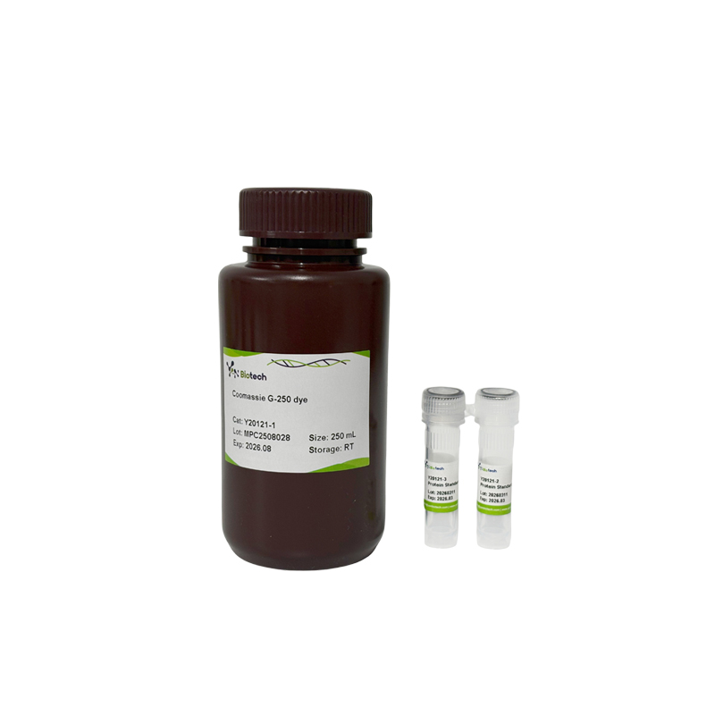
Coomassie (Bradford) Protein Quantitative Assay Kit

Enhance Phosphorylated Protein Stability
Кот. Нет. :
Y20121-250MLБренд :
YanbiotechПроисхождение продукта :
ChinaОбъем :
250mLProduct Information:
| Component Number | Component | Y20121-250ML |
| Y20121-1 | Coomassie G-250 dye | 250 mL |
| Y20121-1 | Bovine Serum Albumin (BSA) Standard Ampules | 25 mg |
| Y20121-1 | Standard Ampules Dilution | 1.5 mL |
Two of the most commonly used methods for quantifying protein concentration are the Bradford method and the BCA method. The principle of the Bradford method is based on the rapid binding of the Coomassie brilliant blue YB360 to proteins. The maximum absorption of Coomassie Brilliant Blue G250 is 488 nm in the free state. After binding to protein, the maximum absorption changed to 595 nm. and the light absorption value is proportional to the content of proteins, so it can be used for the quantitative detection of proteins. This method determines protein concentrations independent of the chemicals in the vast majority of samples, which can be as high as 1 mM for β-mercaptoethanol and 5 mM for dithiothreitol; However, affected by slightly higher concentrations of descaling agents, it is necessary to ensure that the SDS content is less than 0.01%, the Triton X-100 content is less than 0.05%, and the Tween-20, Tween-60, Tween-80, etc. content is less than 0.015%. The BCA Protein Quantitative Detection Kit (Y20122) manufactured by our company is recommended for samples containing descaling agents.
This kit is easy and fast to operate, with high sensitivity and extremely fast detection speed, and can detect protein concentrations ranging from 25-1000 μg/mL.
Storage Conditions:
Product shipped at room temperature. Bovine Serum Albumin (BSA) Standard Ampules stored at 4℃, valid for 12 months. The protein standard solution, prepared with BSA Standard Ampules and Standard Ampules Dilution, store at -20°C and used within 6 months.
Instructions for Use:
1. Preparation of Protein Standard Stock Solution: Dilute 25 mg Bovine Serum Albumin (BSA) Standard Ampules into 1 mL Standard Ampules Dilution; The concentration of Protein standard stock solution is 25 mg/mL, and it should be stored at -20℃.
2. Preparation of Protein Standard Working Solution: An appropriate amount of 25 mg/mL Protein Standard Stock Solution is diluted 50 times with PBS or normal saline to obtain a Protein Standard Working Solution with a final concentration of 0.5 mg/mL. Take care to dilute according to the 10-fold gradient method to ensure accurate dilution.
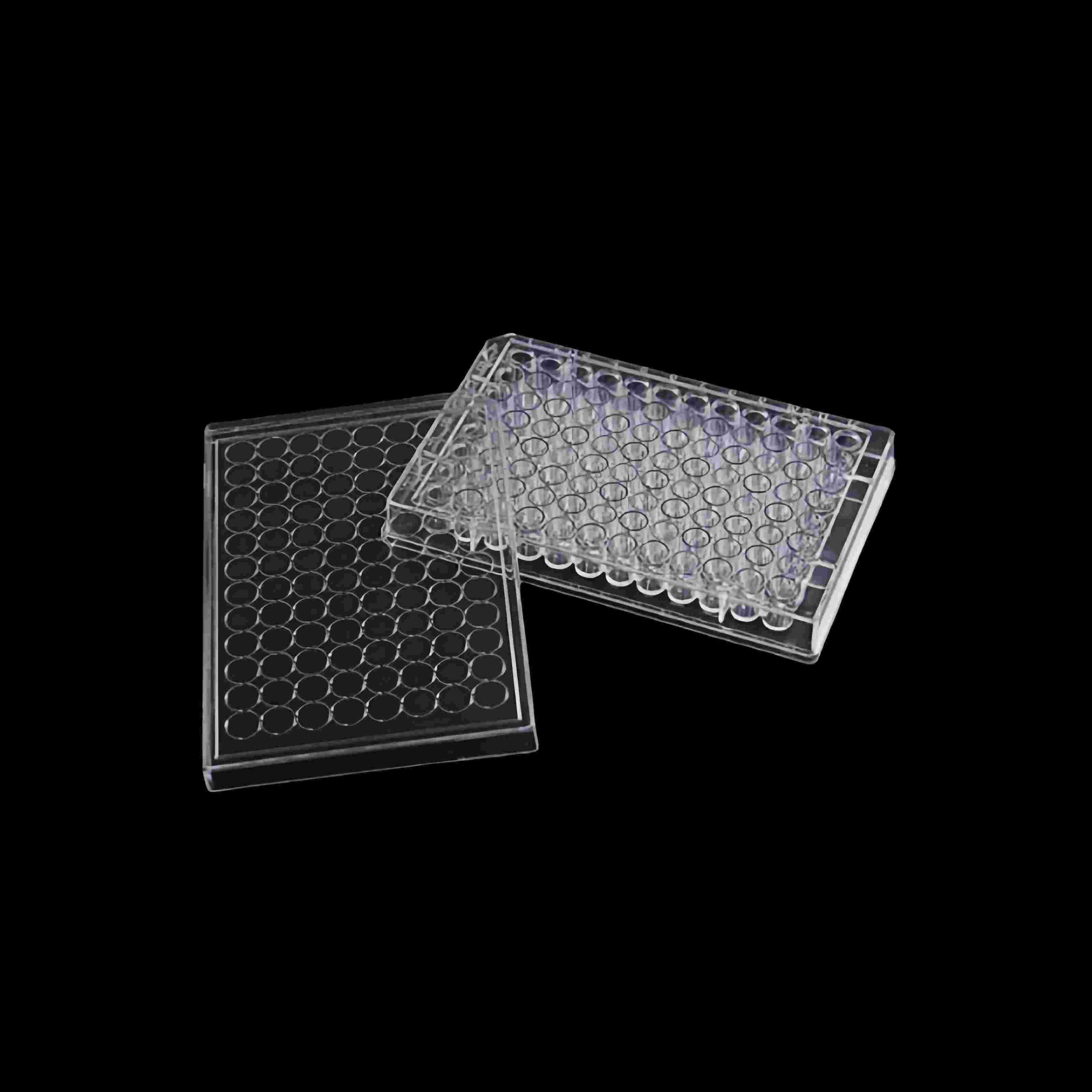
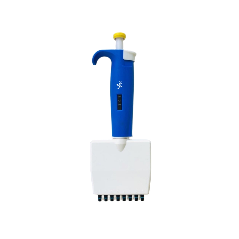
3. Preparation of the standard curve (microplate reader method): The protein standard working solution is added to 96-well plate at 0, 1, 2, 4, 8, 12, 16, 20 μL, and then PBS or normal saline is added to the same 96-well plate in turn at 20, 19, 18, 16, 12, 8, 4, 0 μL to make up the above gradient working solution to 20 μL. The protein concentrations of Gradient curves is 0, 25, 50, 100, 200, 300, 400, and 500 μg/mL.
4. Preparation of the sample: Dilute the sample to be tested appropriately (by pre-experimental detection, the protein concentration of the sample can be within the range of the standard curve to ensure the reliability of the test result). Add 20 μL of samples to a 96-well plate. The sample to be tested and the protein standard are diluted with the same solution.
5. Test Tube Protocol:
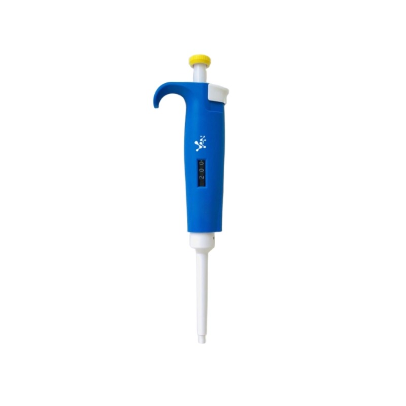
a) Add 200 μL of the Coomassie G250 dye to each tube and mix well (The 96-well plate can be shaken on a vortex for 30 s).
b) Incubate samples for 3-5 minutes at room temperature (RT).
c) The standard curve No. 0 was used as a reference and colorimetrically measured at 595 nm, and the absorbance value of each well was recorded.
6. Calculation:
a)Take the gradient protein content (μg/mL) in the standard curve as the horizontal coordinate and the absorbance value as the vertical coordinate, and plot the standard curve. According to the absorbance value of the measured sample, the protein concentration (μg/mL) of the sample to be tested in the corresponding wells can be found on the standard curve, and then multiplied by the dilution of the sample that is the actual protein concentration of the sample to be tested.
Also: if measured by spectrophotometer, change the gradient standard reaction volume to 1 mL in step 3. Add 0, 0.05, 0.1, 0.2, 0.4, 0.6, 0.8, 1.0 mL of protein standard working solution (concentration of 0.5 mg/mL) to 7 clean glass test tubes or colorimetric tubes, then add 1.0, 0.95, 0.9, 0.8, 0.6, 0.4, 0.2, 0 mL of PBS or saline to each tube in order, and then top up the gradient working solution to 1 mL. The working solution of the gradient was replenished to 1 mL, and each tube was mixed thoroughly to obtain the gradient curve with protein concentrations of 0, 25, 50, 100, 200, 300, 400, 500 μg/mL.
b)Protein samples to be tested were made appropriately diluted and added to new glass test tubes or colorimetric tubes with a sample volume of 1 mL.
c) Add 3 mL of Coomassie G250 dye to each of the above standard curve gradient tubes and sample tubes, mix thoroughly, and let it stand for 3-5 min at room temperature for colorimetric detection by spectrophotometer.
d) The wavelength of the photometer was set at 595 nm during the detection, and the standard curve and the sample to be tested were detected with the standard curve No. 0 tube as the reference for zero adjustment. Plot the standard curve and calculate the protein concentration in the sample to be tested according to the method in step 6.
Notice:
1. Coomassie G250 dye should be brought to room temperature and mixed upside down before use to avoid affecting the sensitivity of the assay.
2. When preparing the protein standard stock solution, make sure that it is well dissolved. It is recommended to dilute the protein standard working solution in a 10-fold gradient, do not dilute 50 times at a time to avoid large errors.
3. Please wear safety glasses, gloves, or protective clothing.
Stay Connected With Us


Авторское право
@ Wuhan Yanbiotech Co., Ltd. Все права защищены
.
 ПОДДЕРЖИВАЕМАЯ СЕТЬ
Xml / политика конфиденциальности
ПОДДЕРЖИВАЕМАЯ СЕТЬ
Xml / политика конфиденциальности
 Русский
 English
 Русский
 Español