Раскрывая невидимое: руководство по системам хемилюминесцентной визуализации
Nov 10, 2025
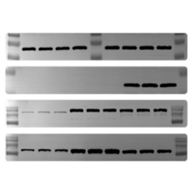
В стремлении разгадать тайны жизни многие из важнейших ответов скрыты на самом видном месте, но они слишком слабы для человеческого глаза. Как нам уловить слабый шёпот света от одного белка? Ответ кроется в силеСистема хемилюминесцентной визуализации.В этом блоге вы узнаете, что представляет собой эта система, почему она так необходима лаборатории и как она ускоряет открытия в области биологических наук.Что такое хемилюминесцентная визуализация?По своей сути система хемилюминесцентной визуализации представляет собой высокочувствительную камеру, предназначенную для обнаружения и документирования света, испускаемого в ходе химических реакций.В лаборатории мы маркируем интересующие нас молекулы (например, белки или ДНК) ферментным репортёром (например, пероксидазой хрена или щелочной фосфатазой). При добавлении субстрата фермент запускает реакцию, в результате которой выделяется свет. Задача системы визуализации — зафиксировать это слабое свечение с предельной точностью и преобразовать его в цифровое изображение, пригодное для количественной оценки.Почему он нужен вашей лаборатории: 3 ключевых преимуществаПочему хемилюминесценция стала золотым стандартом для таких методов, как вестерн-блоттинг? Преимущества очевидны:1. Крайняя чувствительностьОн способен обнаруживать целевые белки вплоть до фемтограммового уровня, что значительно превосходит традиционные колориметрические методы и незаменимо для малораспространенных мишеней.2. Широкий динамический диапазонОн может точно улавливать как очень сильные, так и очень слабые сигналы за одну экспозицию, обеспечивая точную количественную оценку в широком диапазоне уровней экспрессии.3. Простота и надежностьПоскольку метод не требует внешнего источника света, он позволяет избежать таких проблем, как рассеяние света и фотообесцвечивание. Это обеспечивает более чистые данные с низким фоном и высокой воспроизводимостью.Основные приложения: где все проявляется?Эта система — универсальная «рабочая лошадка» в любой лаборатории молекулярной биологии:Вестерн-блоттинг: Типичное применение. Для обнаружения специфических белков с высокой специфичностью и чувствительностью.Саузерн/Нозерн-блоттинг: Для обнаружения специфических последовательностей ДНК и РНК.Гибридизация колоний/бляшек: Быстро проведите скрининг бактериальных колоний или фаговых бляшек на предмет интересующего вас гена.Анализы репортерных генов: Изучите активность промотора гена, измеряя выход фермента люциферазы.Дот-блоты и белковые массивы: Высокопроизводительный метод одновременного анализа нескольких образцов.Выбор правильной системы для ваших нуждНе все устройства для обработки изображений одинаковы. Вот что следует учитывать:1. Чувствительность: Самая важная характеристика. Обратите внимание на низкий предел обнаружения.2. Качество камеры: Для получения изображений с низким уровнем шума решающее значение имеет высококачественная охлаждаемая ПЗС- или sCMOS-камера.3. Динамический диапазон: Гарантирует, что вы не пропустите слабые полосы рядом с сильными.4. Программное обеспечение: Необходимо иметь удобное программное обеспечение с мощными инструментами для сбора данных, количественной оценки (например, анализа плотности полос) и создания отчетов.5. Автоматизация: Для основных объектов или лабораторий с высокой производительностью обратите внимание на такие функции, как автоматическая фокусировка и загрузка нескольких образцов.


 ПОДДЕРЖИВАЕМАЯ СЕТЬ
Xml / политика конфиденциальности
ПОДДЕРЖИВАЕМАЯ СЕТЬ
Xml / политика конфиденциальности Русский
 English
 Русский
 Español